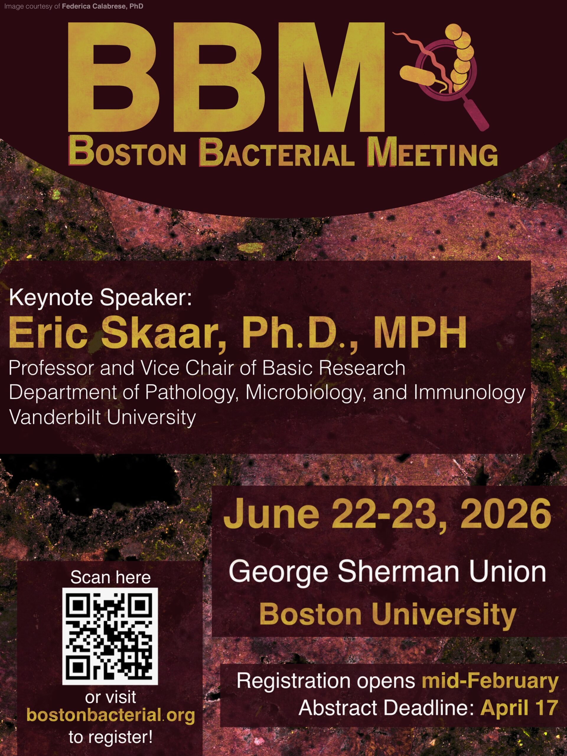
The micrograph featured on the BBM 2026 posterwas taken by Federica Calabrese, a postdoc in Jeffrey Marlow’s lab at Boston University. The image shows a mineral chip from a serpentinite rock colonized with endemic microbial communities after two months of in situ incubation at The Cedars in Northern California. The chip’s surface is outlined by different colors (a merge of blue, magenta, grey, and yellow) that represent the diverse autofluorescence wavelengths emitted by the minerals composing the rock. Microbial cells labeled with SYBR Gold, a fluorescent DNA stain, are shown in green; they colonized just a few spots within the surface of the rock chip.
The poster was designed by Ashley Luo (Tufts University) and Lauren Frost (Northeastern University), both graduate students and members of the BBM organizing committee.
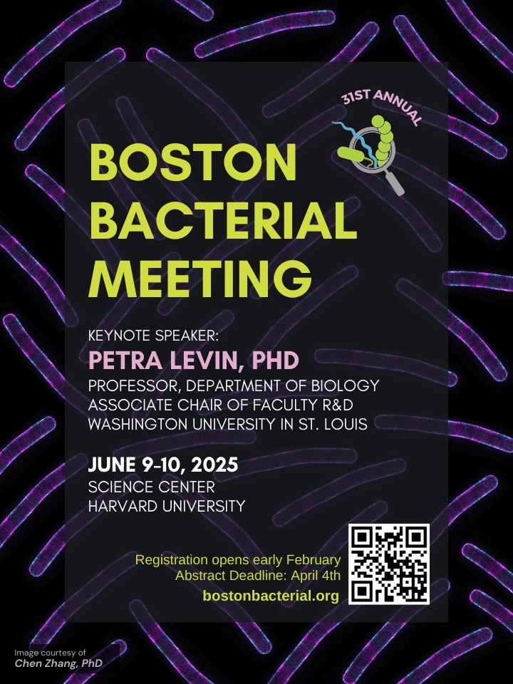
BBM 2025 Poster
The micrograph featured on the BBM 2025 posterwas taken by Chen Zhang, a postdoc in David Rudner’s lab at Harvard Medical School. The image shows the localization of different S-layer proteins in Bacillus anthracis.
The poster was designed by Abigail Rivera Seda, a BBM organizing committee member and graduate student at Tufts University.
BBM 2024 Poster
The micrograph featured on the BBM 2024 posterwas taken by Andrea Vettiger back when he was a postdoc in Tom Bernhardt’s lab at Harvard Medical School; he is currently an Assistant Professor at the University of Lausanne. The image shows a composite of the outer membrane protein Pal (magenta) and bacterial cytoskeletal element MreB (white) in Escherichia coli. The image was acquired using a combination of structured illumination and total internal reflection microscopy. MreB displacement was analyzed by TrackMate (colors).
The poster was designed by Katherine Suarez, a BBM organizing committee member and graduate student at Harvard Medical School.
Website Images
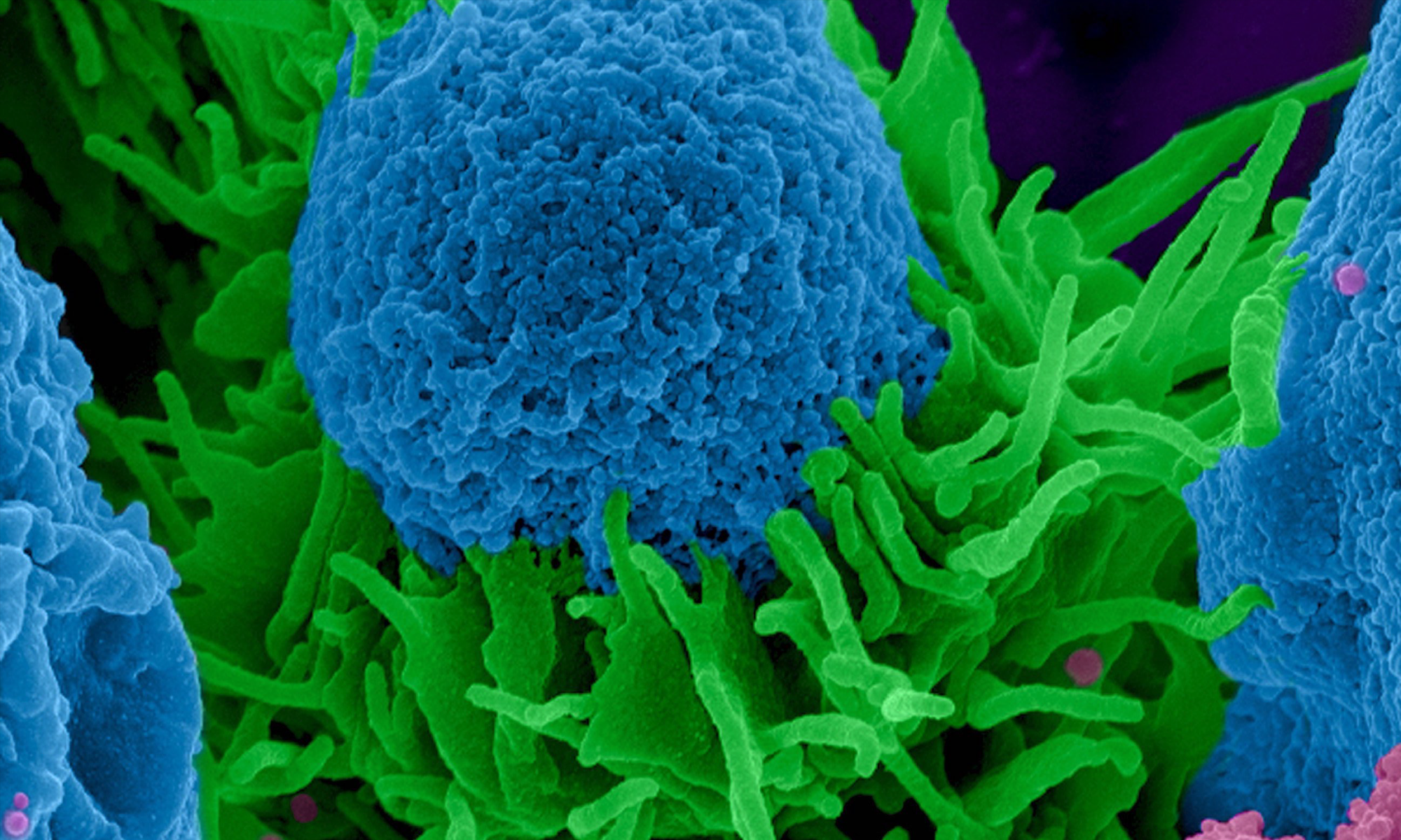
This scanning electron micrograph showing mouse macrophages (green) interacting with Cryptococcus neoformans (blue) was provided by Sabriya Stukes and Hillary Guzik of the Albert Einstein College of Medicine.
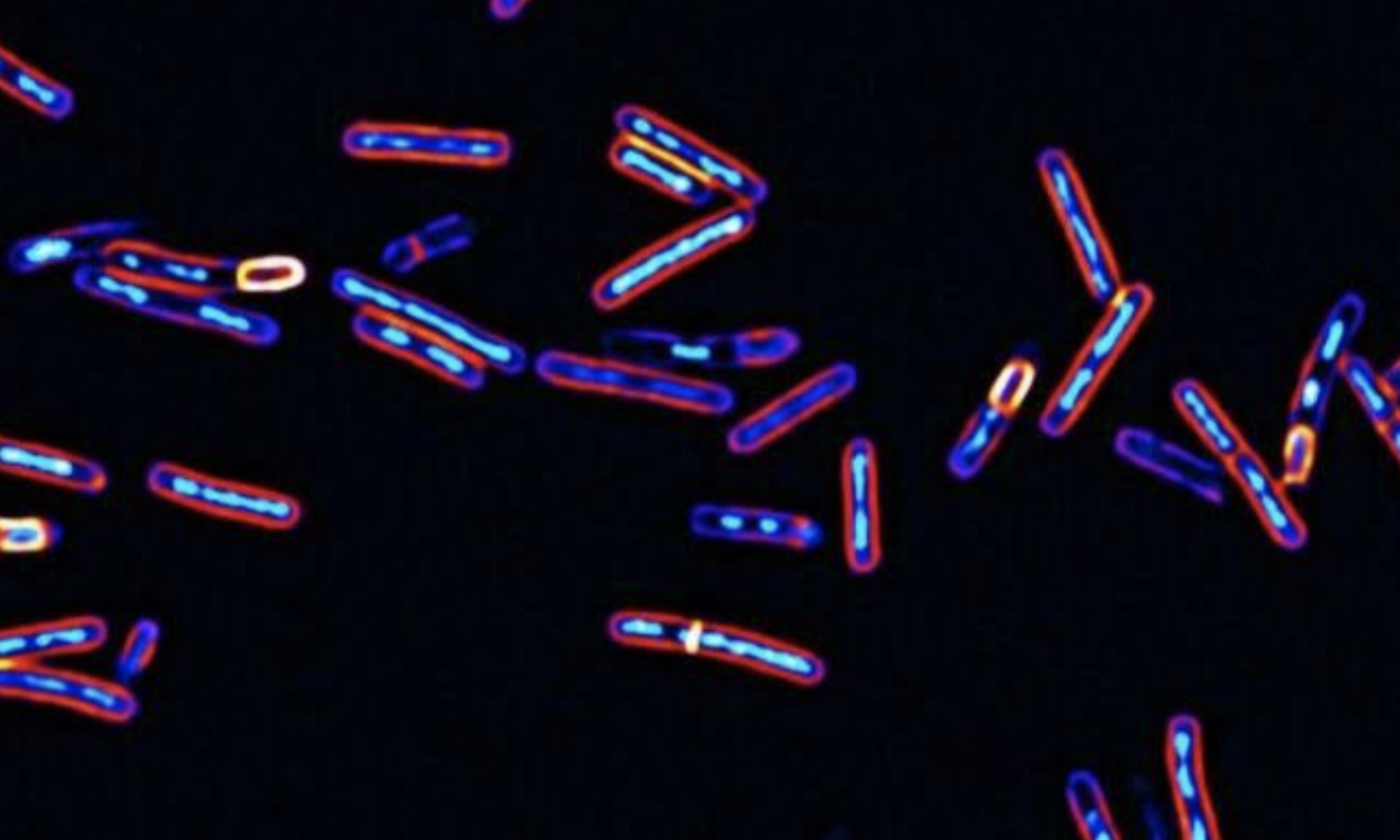
This image showing sporulating Clostridioides difficile cells stained with DNA and membrane dyes was provided by John Ribis of Tufts University.
This image obtained from a confocal microscope shows the spatial organization of a microbial consortium from the biofilm of a human tongue. It revealsa dense community of bacteria (blue, cyan, magenta, and yellow) organized around a core of host epithelial cells (pink/white). The micrograph was provided by Tabita Ramirez-Puebla of the Forsyth Institute.